
Exfoliative Cell DNA Quantitative Analysis System
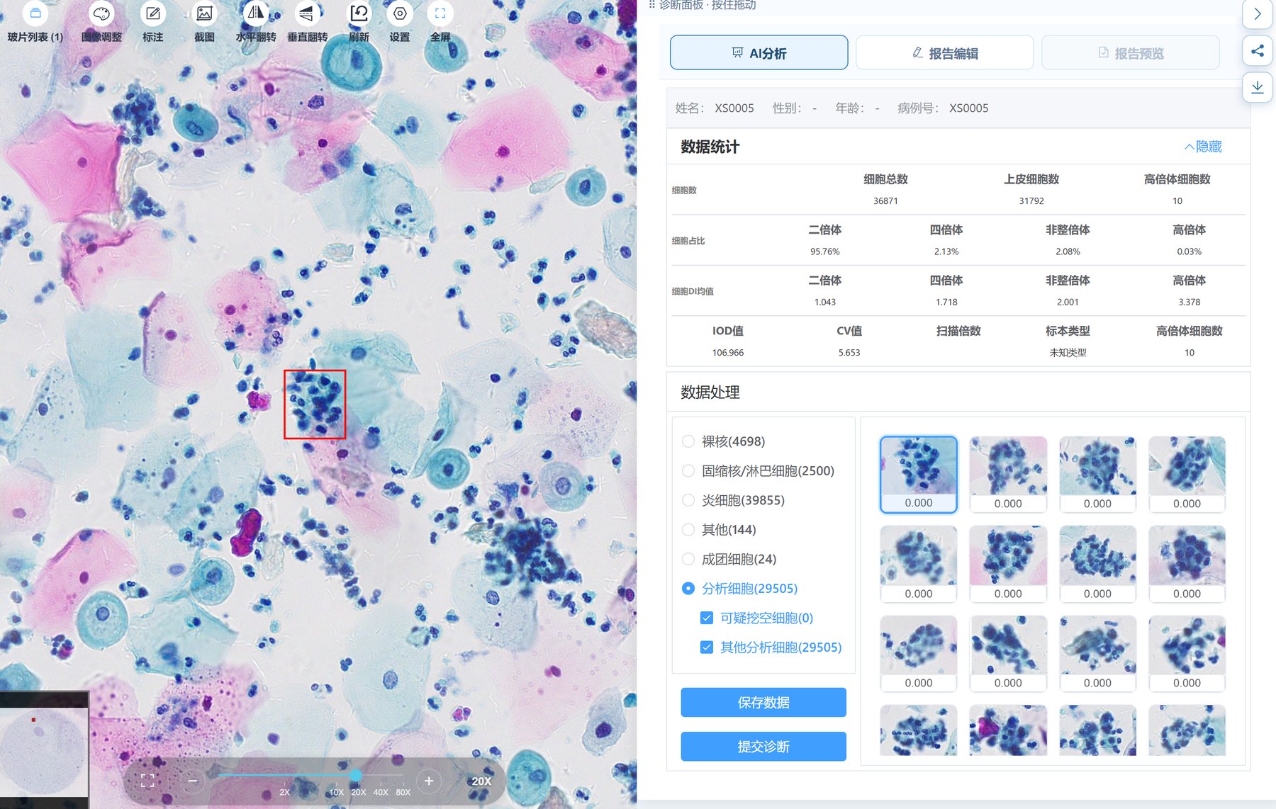
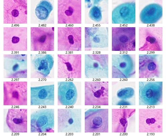
The system uses software to control a fully automated scanning platform and digital camera, automatically scanning and statistically analyzing compound-stained cervical exfoliative cell smears. It measures the DNA content in the nuclei of all cells in the sample to determine whether precancerous lesions have occurred.
Core Features
Early Detection of Cancer and Precancerous Lesions
Precisely measures nuclear DNA content to sensitively detect cancer and precancerous lesions, of great value in cervical cancer screening.
Highly Predictive
When cells become malignant, DNA content changes appear 3–5 years earlier than morphological changes.
Fully Automated Scanning
Software controls the scanning platform and camera to automatically scan smears and capture cells.
Histograms & Scatter Plots
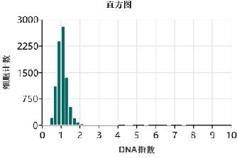
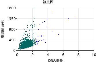
Automatically generates DNA index histograms and scatter plots, making diploid, tetraploid and aneuploid cells clear at a glance.
Cell Classification & Counting
Naked nuclei, lymphocytes, inflammatory cells, cell clusters and more are classified automatically; DNA content is calculated only for analyzed cells.
Quality Control Metrics
Outputs QC metrics such as IOD and CV values to ensure reliable results.
Histograms and Scatter Plots Show Ploidy Distribution at a Glance
The system calculates the DNA index (DI) of every analyzed cell, automatically distinguishes diploid, tetraploid, aneuploid and hyperploid cells, and provides QC metrics such as IOD and CV values, offering a quantitative basis for assessing precancerous risk.
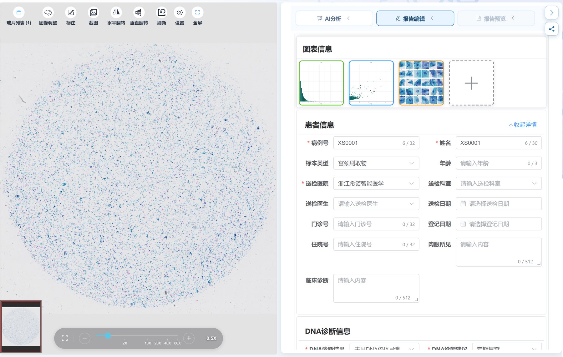
Product Interface & Photos
Workflow
Smear Preparation
Compound-stained cervical exfoliative cell smears
Automated Scanning
Fully automated scanning and nucleus capture
DNA Measurement
DNA index calculated for each nucleus
Report Output
Histograms, scatter plots and diagnostic suggestions
Related Products
 H&E Histology
Histopathology AI
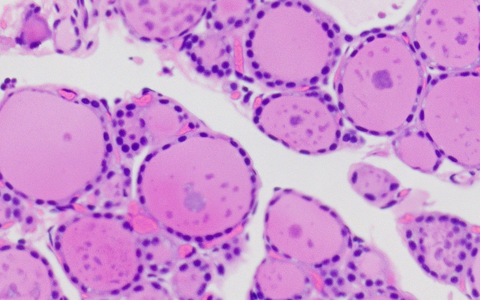
For H&E histopathology slides, providing whole-slide pre-screening and suspicious region localization, from whole-slide survey to case-level conclusions, with evidence traceable throughout.
Learn More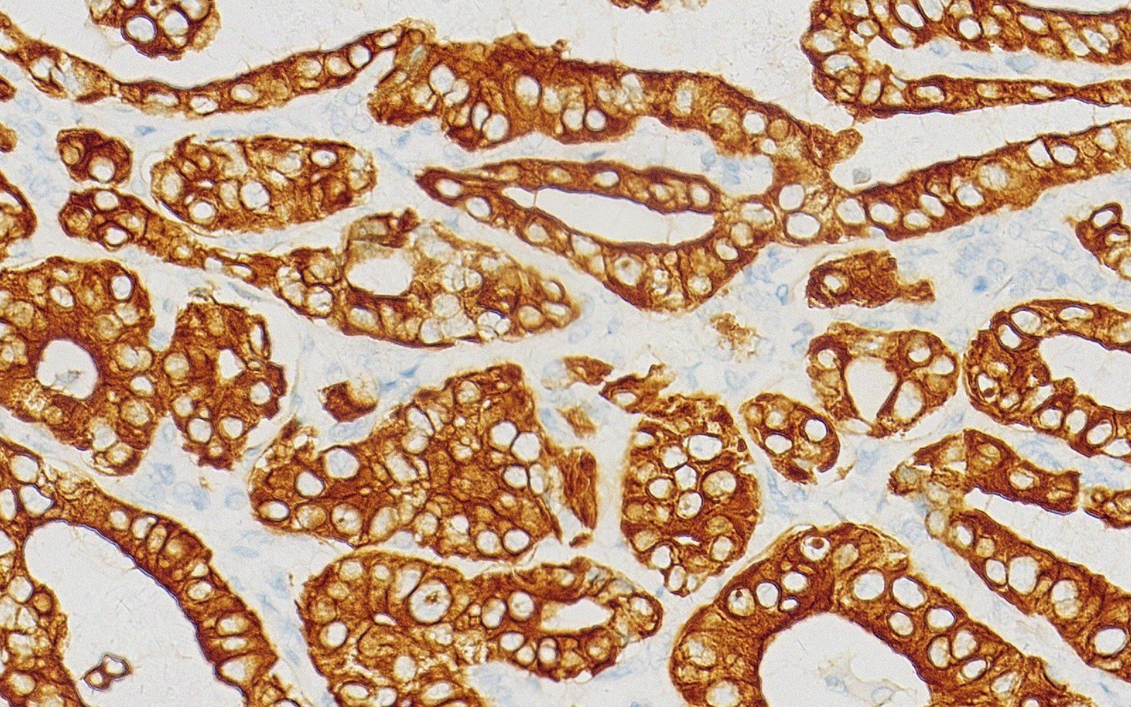 IHC
IHC AI
Assists analysis of stained regions and cellular expression features in IHC slides, with intuitive markings to support pathologist interpretation and review.
Learn More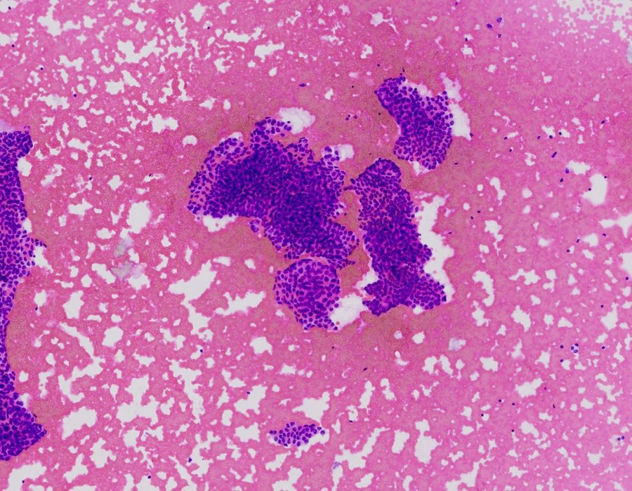 FNA Cytology
 AI
Segments and interprets nuclei and cell clusters one by one with evidence, providing risk stratification and Bethesda classification, with conclusions traceable to specific cells for verification.
Learn More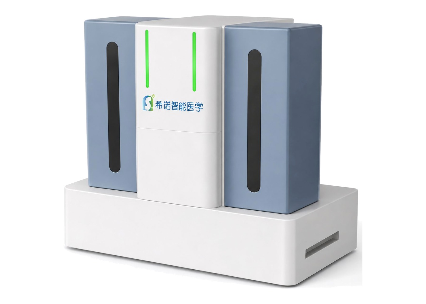
Digital Pathology Scanner
Throughput configurations from 200 to over 400 slides, 20× / 40× high-definition scanning; digital slides connect directly to AI-Assisted Diagnosis, serving as the data entry point of the AI pathology loop.
Learn MoreBring AI Agents to Your Department
From digital scanning to AI-assisted diagnosis, Xinuo Intelligent Medical provides standardized, deployable end-to-end AI pathology solutions.
 Scan to follow Xinuo Intelligent Medical