
FuxiMind
Pathology-Native AI Multimodal Foundation Model
From recognizing an image to actively seeking evidence. FuxiMind is our in-house, pathology-native multimodal foundation model, providing intelligent pre-screening and AI-assisted diagnosis for whole slide images, with every conclusion traceable and verifiable.
Not Just a Classifier, but an AI That Finds Evidence
Conventional pathology AI typically outputs labels for a single task and a single field of view; FuxiMind works across the whole slide and the whole case, thinking like a pathologist.
Whole-Slide Multi-Scale Understanding
From low-power tissue architecture to high-power cell morphology, it surveys and localizes at the WSI level.
Active Evidence-Seeking
Automatically finds and numbers (CE) the key evidence regions supporting the diagnosis, with each conclusion mapped to its evidence.
Case-Level Reasoning
Interprets multiple slides from the same case together, outputting a diagnostic conclusion and a detailed comprehensive diagnosis.
Multimodal Fusion
Fuses pathology images, clinical information, and report text to generate structured text that pathologists can cite directly.
Multiple Specimen Types
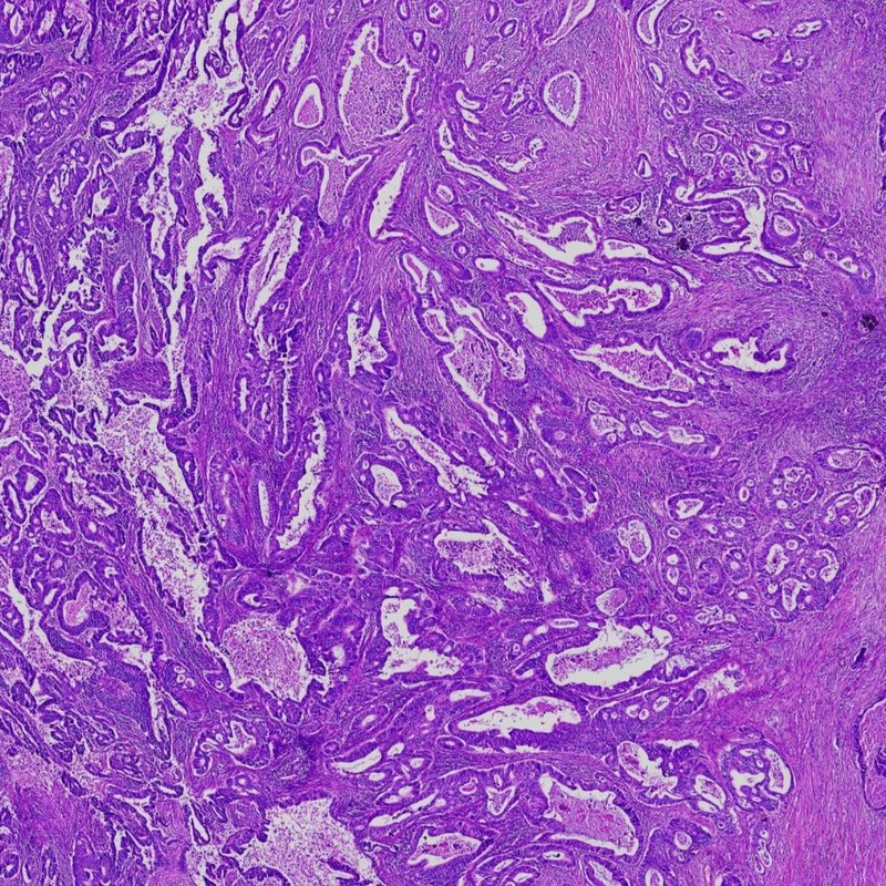
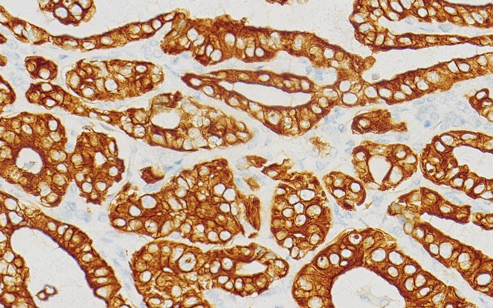
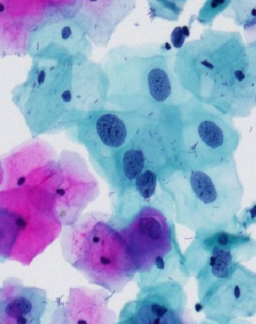
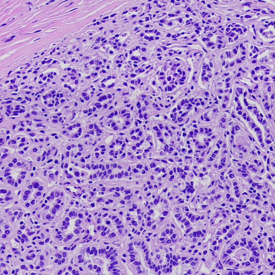
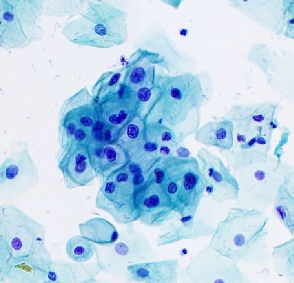
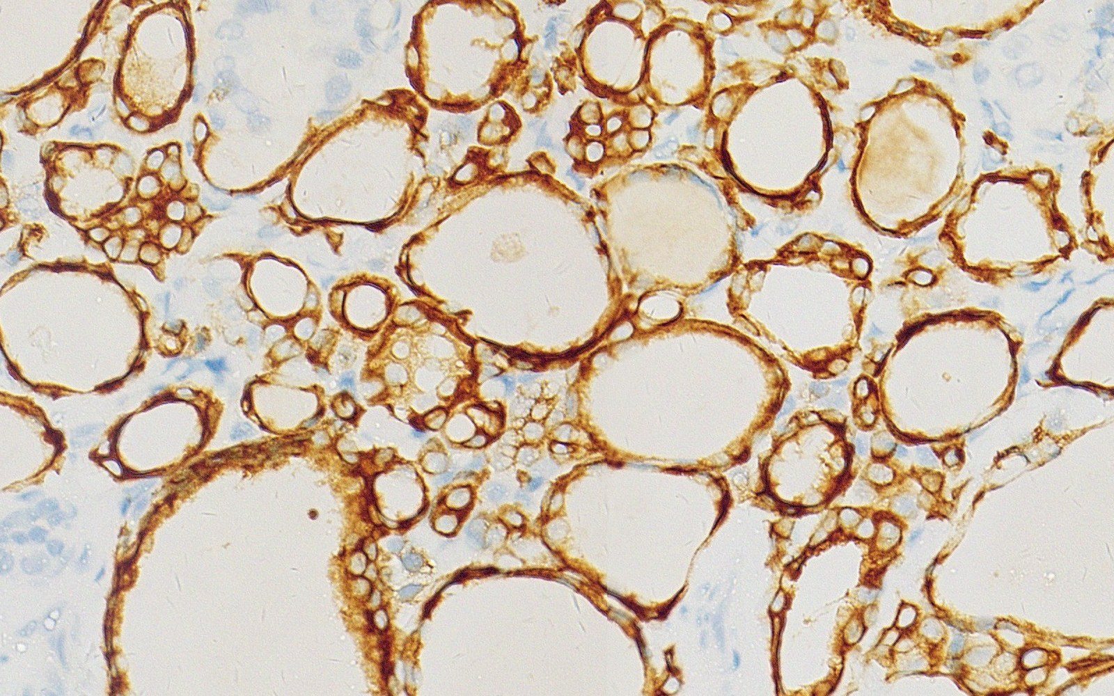
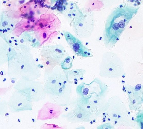
One foundation supports histopathology, IHC, FNA cytology, exfoliative cytology, DNA ploidy, and more.
Continuous Learning
Pathologist review feedback flows back, and the model keeps improving with in-hospital data, forming an AI-native closed loop.
A Layered Foundation for Pathology AI
Clinical Application Layer
Creating value across clinical scenarios
Model Layer
FuxiMind Pathology-Native AI Multimodal Foundation Model
Data Layer
Diverse, large-scale, high-quality
The Evidence Chain of a Real Case
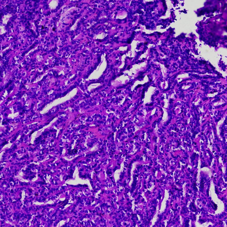
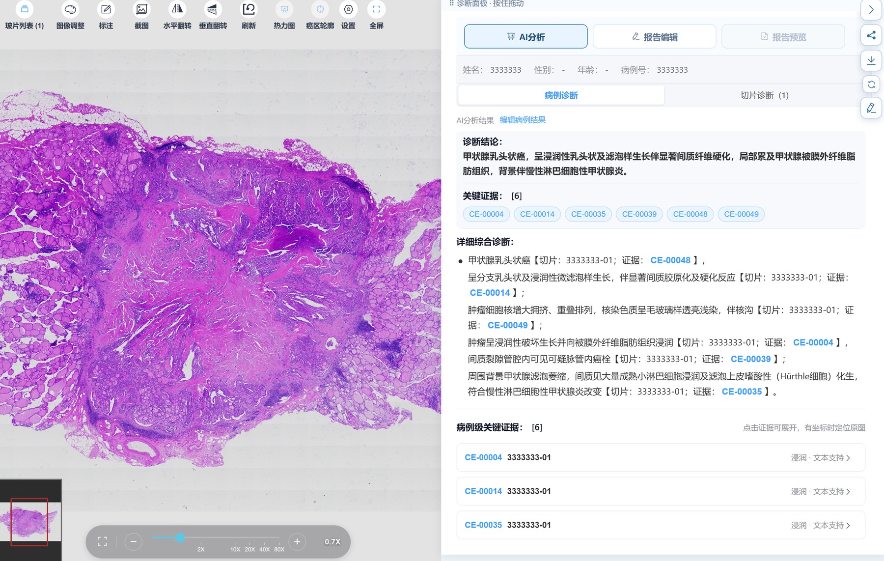
Below is the actual platform interface: FuxiMind interprets the whole slide of thyroid case 3333333 and returns 6 pieces of case-level key evidence.
Diagnostic Conclusion
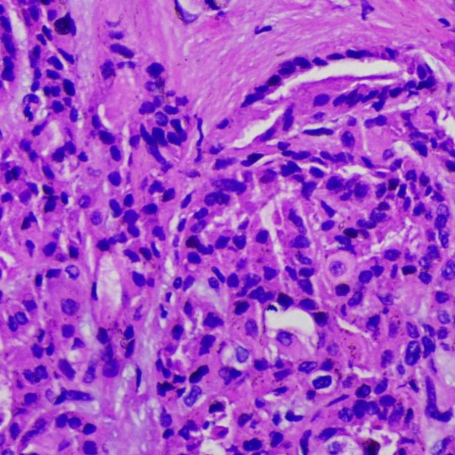
Papillary thyroid carcinoma with invasive papillary and follicular growth and marked stromal fibrosis, focally extending into extrathyroidal fibroadipose tissue, on a background of chronic lymphocytic thyroiditis.
Margin · Text Support
Key region supporting "papillary thyroid carcinoma".
Infiltration · Text Support
Enlarged, crowded and overlapping tumor nuclei with ground-glass, pale chromatin and nuclear grooves.
Infiltration · Text Support
Destructive invasive tumor growth with infiltration into extracapsular fibroadipose tissue.
Cytology, Equally Evidence-Based
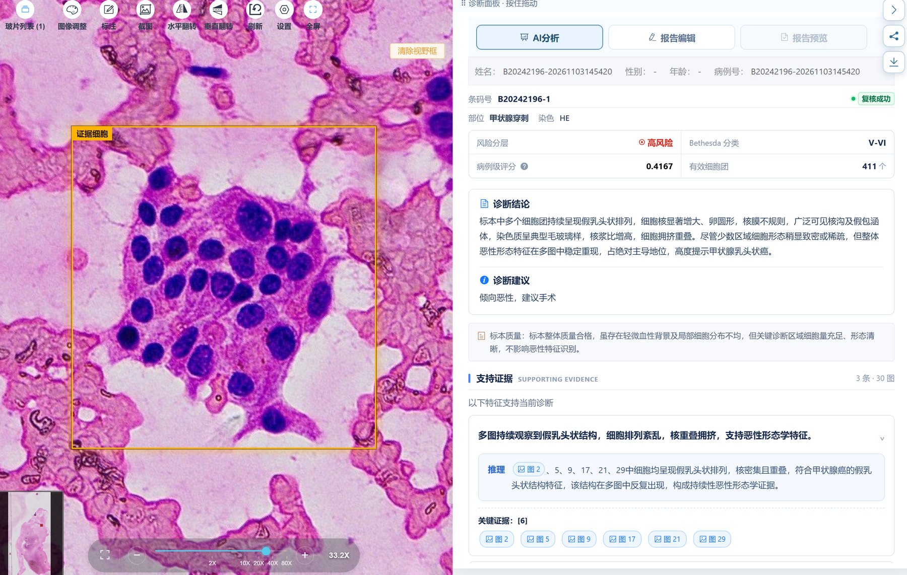
In FNA cytology, FuxiMind provides risk stratification, a case-level score, and Bethesda classification, lists the morphological features and reasoning supporting the current diagnosis, and attaches all key evidence images for pathologists to check one by one.
Risk Stratification · Bethesda Classification
Case-level scoring and classification suggestions, with concurrent specimen adequacy assessment.
Reasoning + Evidence Images
Every morphological feature comes with a reasoning note and evidence image number.
Bring FuxiMind to Your Pathology Department
Supports on-premises deployment and integrates with in-hospital LIS / PIS workflows.
 Scan to follow Xinuo Intelligent Medical