
FNA Cytology AI-Assisted Diagnosis System
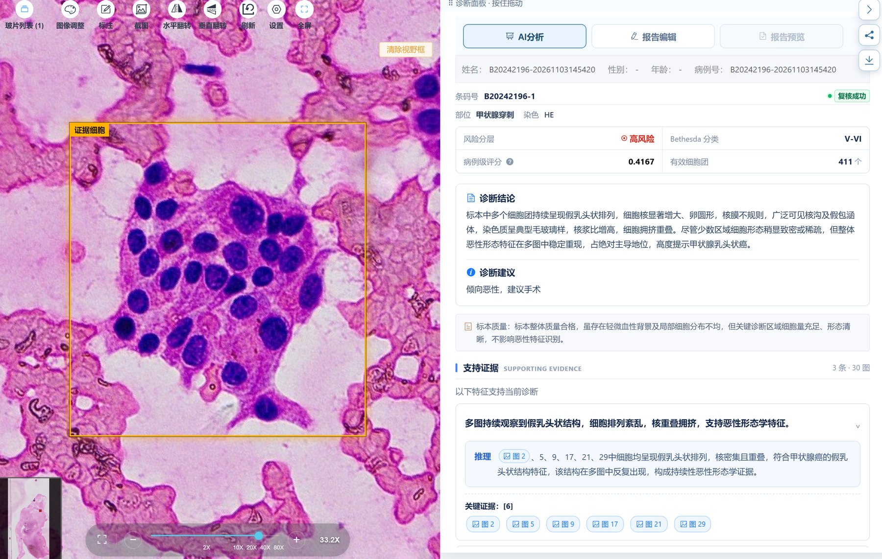
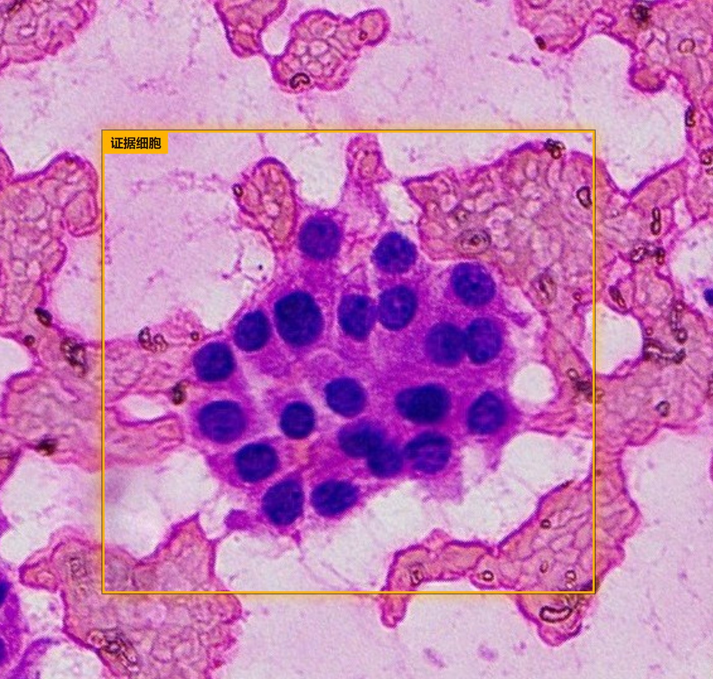
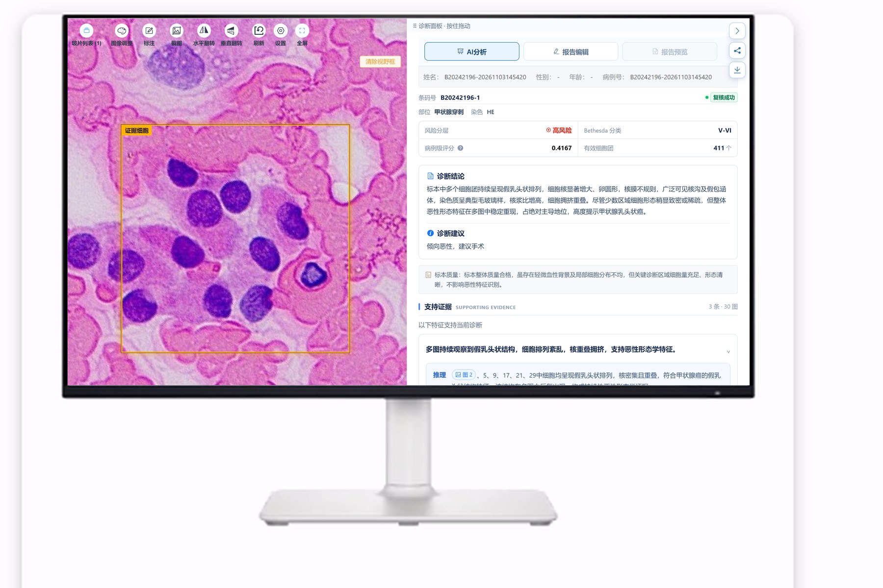
Designed for fine-needle aspiration cytology images, the system helps identify cell cluster distribution and nuclear morphology, quickly marking cell regions that need attention. It supports segmentation and evidence-based interpretation of individual nuclei and cell clusters, and every conclusion can be traced back to specific nuclei for verification.
Core Features
Cell Cluster & Nuclear Feature Analysis
Segments individual nuclei and cell clusters and analyzes morphological features such as nuclear enlargement, nuclear grooves and intranuclear pseudoinclusions.
Suspicious Cell Region Localization
Automatically locates suspicious cell regions and quickly marks fields that need attention.
Risk Stratification & Bethesda Classification
Provides case-level scores, risk stratification and Bethesda classification suggestions.
Key Fields & Evidence Retention
Every piece of supporting evidence comes with images and reasoning, traceable back to specific cells.
Specimen Adequacy Assessment
Flags insufficient cellularity, blood contamination and similar issues to help determine specimen adequacy.
Diagnostic Conclusions & Recommendations
Outputs diagnostic conclusions and recommendations, signed out after physician review.
Product Interface & Photos
Workflow
Smear Scanning
Whole-slide digitization of aspiration smears
Cell Segmentation
Individual segmentation of nuclei and cell clusters
Evidence-Based Interpretation
Risk stratification and supporting evidence
Review & Sign-Out
Signed out after image-by-image physician review
Related Products
 H&E Histology
Histopathology AI
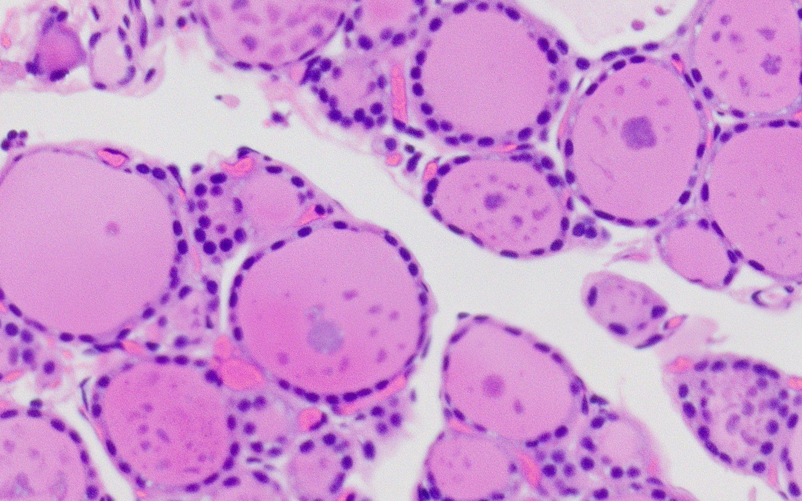
For H&E histopathology slides, providing whole-slide pre-screening and suspicious region localization, from whole-slide survey to case-level conclusions, with evidence traceable throughout.
Learn More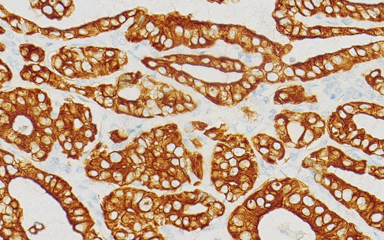 IHC
IHC AI
Assists analysis of stained regions and cellular expression features in IHC slides, with intuitive markings to support pathologist interpretation and review.
Learn More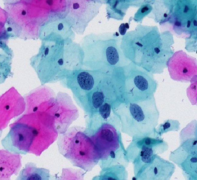 Cervical TBS
Exfoliative Cytology AI
Performs morphological classification and grading of cervical exfoliative cells and issues structured diagnostic reports under the TBS system.
Learn More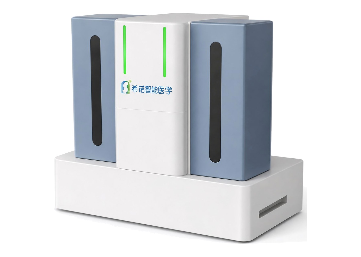
Digital Pathology Scanner
Throughput configurations from 200 to over 400 slides, 20× / 40× high-definition scanning; digital slides connect directly to AI-Assisted Diagnosis, serving as the data entry point of the AI pathology loop.
Learn MoreBring AI Agents to Your Department
From digital scanning to AI-assisted diagnosis, Xinuo Intelligent Medical provides standardized, deployable end-to-end AI pathology solutions.
 Scan to follow Xinuo Intelligent Medical